Complete The Labeling Of The Diagram Of The Upper Respiratory Structures
Frontal sinus cribriform plate of ethmoid bone. Complete the labeling of the diagram of the upper respiratory structures sagittal section.
 Structure Of The Human Respiratory System Explicated With Diagrams
Fill in the missing organs of the respiratory system.

Complete the labeling of the diagram of the upper respiratory structures. Which lymphatic structure drains lymph from the right upper limb and. Name lab timedate anatomy of the respiratory system upper and lower respiratory system structures 1. Complete the labeling of the diagram of the upper respiratory structures sagittal section.
Cribriform plate of ethmoid bone 7. Two pairs of vocal folds are found in the larynx. Anatomy of blood vessels conduction system of the heart electrocardiography human cardiovascular physiology.
Blood pressure pulse determinations anatomy of the respiratory system respiratory system physiology. Complete the labeling of the diagram of the upper respiratory structures sagittal section. Respiratory assignment bio 169 respiratory system.
Learn vocabulary terms and more with flashcards games and other study tools. Anatomy of the exercise36 respiratory system. They are upper respiratory tract and lower respiratory tract.
Sphenoidal sinus opening of auditory tube frontal sinus 43me rr. 53 if 3an c t. Review sheet 36 283.
Nose and mouth air enters nasal cavity larynx both air and food move through trachea bronchi large tubes leading to both lungs lungs. Complete the labeling of the diagram of the upper respiratory structures sagittal section. Due to respiratory movement 50.
Upper and lower respiratory system structures. A p ii lab practical 2 review. Anatomy of the respiratory system upper and lower respiratory system structures 1.
Step 1 of 5 the respiratory tract is divided into two parts. Start studying ap ii review sheet 36 anatomy of the respiratory system. Complete the labeling of the diagram of the upper respiratory structures sagittal section.
In the diagram at left which of the labeled structures is the thoracic duct. Complete the labeling of the diagram of the upper respiratory structures sagittal section. Anatomy of the respiratory system flashcards and study them anytime anywhere.
Anatomy Of The Respiratory System
 Structure Of The Human Respiratory System Explicated With Diagrams
 Human Skeletal System Parts Functions Diagram Facts
 Structure Of The Human Respiratory System Explicated With Diagrams
 Pdf Respiratory Control By Ventral Surface Chemoreceptor Neurons In
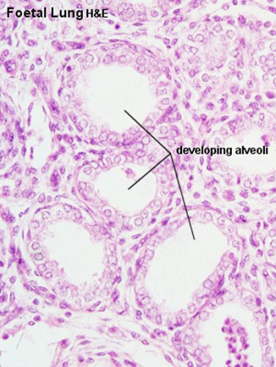
 Respiratory System Histology Embryology
 Your Lungs Respiratory System For Kids Kidshealth
 Human Respiratory System Description Parts Function Facts
 Lungs Human Anatomy Picture Function Definition Conditions
 Fascinating And Amazing Human Body Facts And Trivia Disabled World

 What Is An Organ System Definition Pictures Video Lesson
 Lung Anatomy Overview Gross Anatomy Microscopic Anatomy
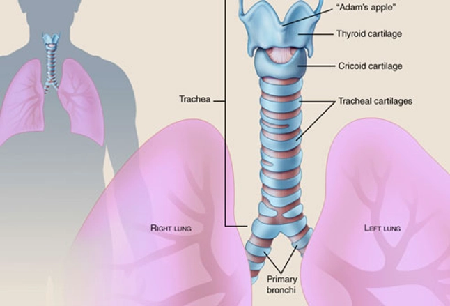
 The Trachea Human Anatomy Picture Function Conditions And More
 Ciliated Epithelium Function Structure Diagram Video Lesson
Brain Structure And Function Brain Injury British Columbia
 Diaphragm Anatomy Function Diagram Conditions And Symptoms
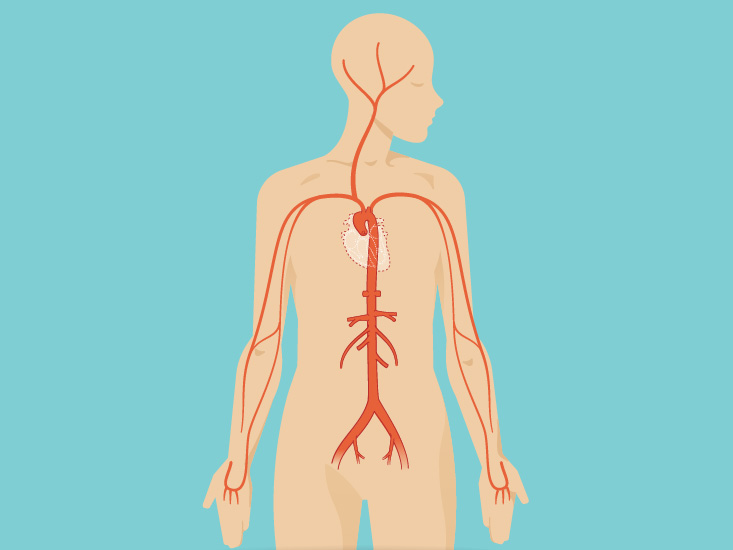
 This Diagrams Shows The Major Arteries In The Human Body School


0 Response to "Complete The Labeling Of The Diagram Of The Upper Respiratory Structures"
Post a Comment