The Diagram Below Shows A Double Stranded Dna Molecule Parental Duplex
Drag the correct labels to the appropriate locations in the diagram to show the composition of the daughter dna molecules after one and two cycles of dna replication. The parental dna is shown in dark blue the newly synthesized dna is light blue and the rna primers associated with each strand are red.
 Rara Binds Double Stranded Dna In The Presence Of Atp S And
Show transcribed image text the diagram below shows a bacterial replication fork and its principal proteins.

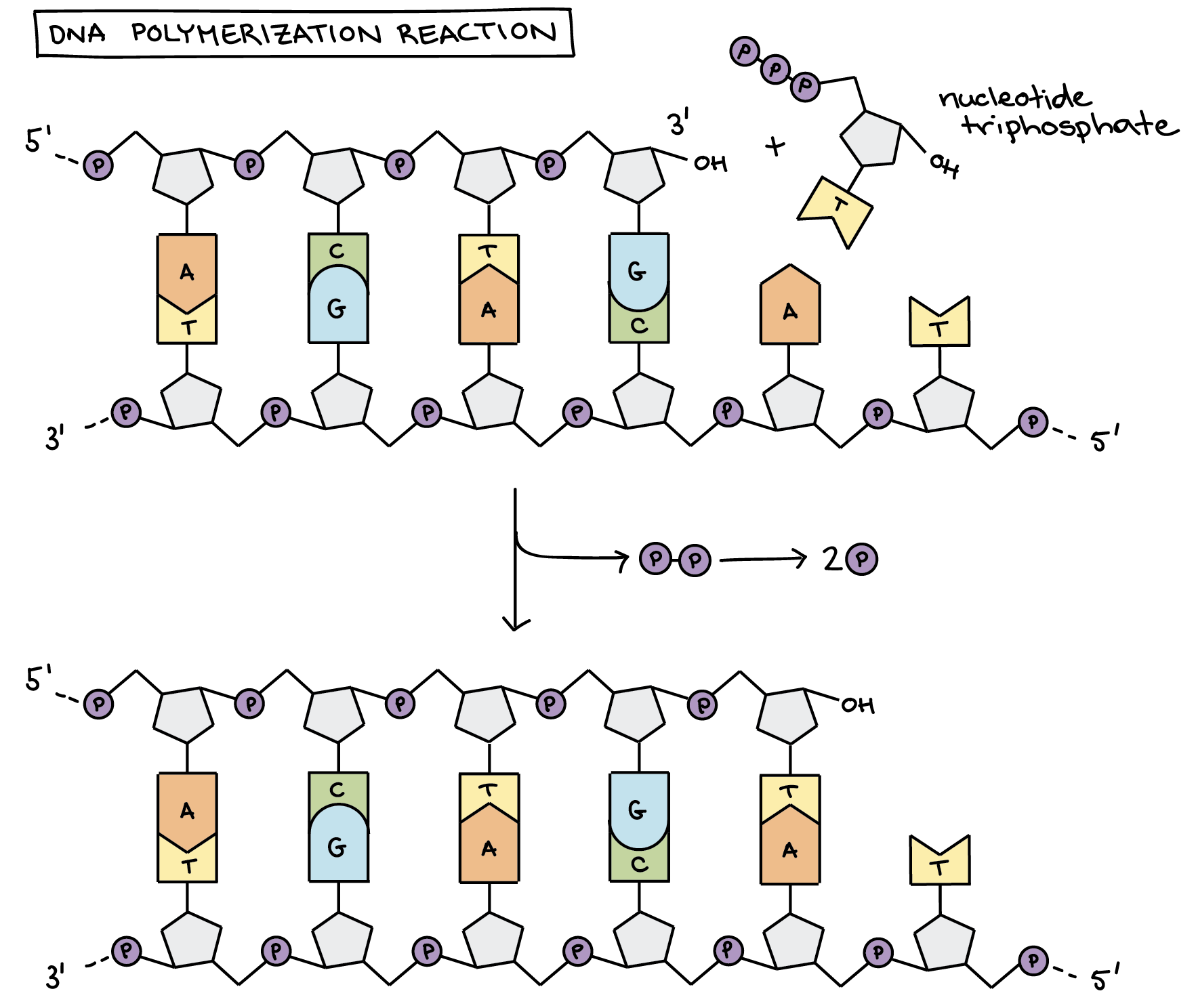
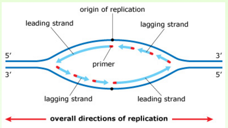
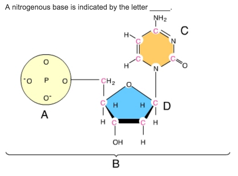
The diagram below shows a double stranded dna molecule parental duplex. Nucleic acids are made up of chains of many repeating units called nucleotides see bottom left of figure 1 below. In the labels the original parental dna is blue and the dna synthesized during replication is red. The diagram below shows a replication bubble with synthesis of the leading and lagging strands on both sides of the bubble.
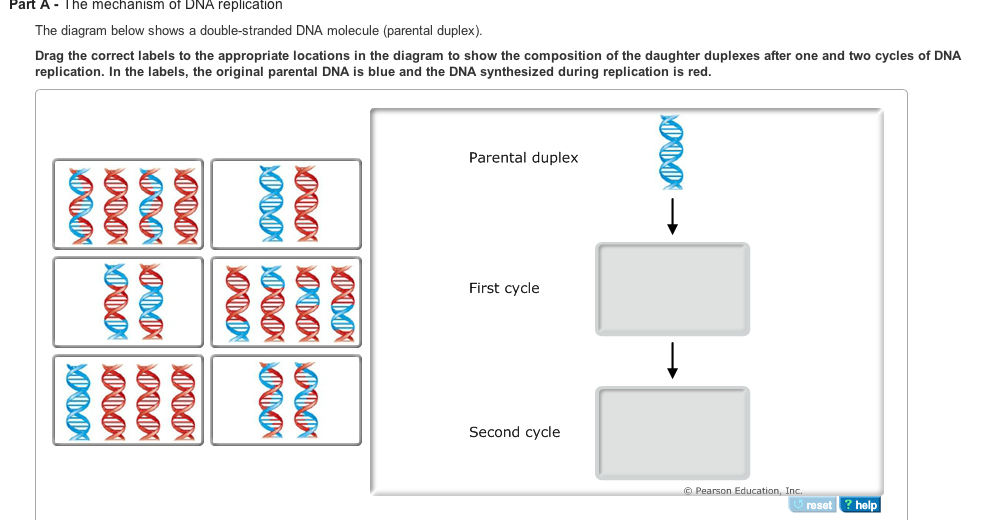
Drag the correct labels to the appropriate locations in the diagram to show the composition of the daughter duplexes after one and two cycles of dna replication. The structure of dna. The dna molecule actually consists of two such chains that spiral around an imaginary axis to form a double helix spiral.
In the labels the original parental dna is blue and the dna synthesized during replication is red. The diagram below shows a double stranded dna molecule parental duplex. Use pink labels for the pink targets and blue labels for the blue targets.
In the labels the original parental dna is blue and the dna synthesized during replication is red. The diagram below shows a double stranded dna molecule parental duplex. The diagram below shows a double stranded dna molecule parental duplex.
In the labels the original parental dna is blue and the dna synthesized during replication is red. The diagram below shows a replication bubble with synthesis of the leading and lagging strands on both sides of the bubble. The parental dna is shown in dark blue the newly synthesized dna is light blue and the rna primers associated with each strand are red.
Drag the correct labels to the appropriate locations in the diagram to show the composition of the daughter duplexes after one and two cycles of dna replication. Part a the mechanism of dna replication the diagram below shows a double stranded dna molecule parental dna. Drag the correct labels to the appropriate locations in the diagram to show the composition of the daughter duplexes after one and two cycles of dna replication.
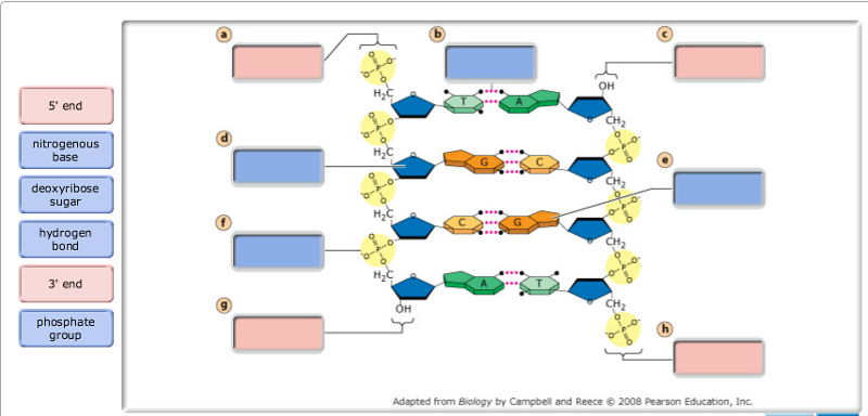
Drag the labels to their appropriate locations in the diagram to describe the name or function of each structure. The origin of replication is indicated by the black dots on the parental strands. The origin of replication is indicated by the black dots on the parental strands.
 Effects Of Homology Length And Superhelicity On Paranemic Joint
 Molecular Mechanism Of Dna Replication Article Khan Academy
 Architecture Of The E Coli Replication Fork The Parental Duplex
 Exam 3 Chs 5 Dna Structure And Replication Machinery 16 The
 Mechanisms Of Maintenance Of Genome Stability The Original Parental
 Nucleic Acid Chemical Compound Britannica Com
Iii Recombination Lewin Chpt 33
 Exam 3 Chs 5 Dna Structure And Replication Machinery 16 The
 Mastering Biology Chapter 16 Rhs Homework
 Comparison Of Single Stranded And Double Stranded Dna Binding By
 Bach1 Is Preferentially Sequestered On Forked Duplex Dna Molecules
 Solved Dna Replication Is The Mechanism By Which Dna Is C
 Exam 3 Chs 5 Dna Structure And Replication Machinery 16 The


Hello I am so delighted I located your blog, I really located you by mistake, while I was watching on google for something else, Anyways I am here now and could just like to say thank for a tremendous post and a all round entertaining website. Please do keep up the great work. phone tracker app
ReplyDelete