Identify The Structures Labeled A B And C In The Diagram Of A Sarcomere Above
Eany of the above can produce an action potential in the muscle cell. The brain stem connects the brain with the the brain stem connects the brain to the spinal cord.
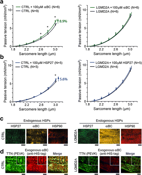 Translocation Of Molecular Chaperones To The Titin Springs Is Common
A acetylcholine binds to chemically gated channels in the end plate membrane.

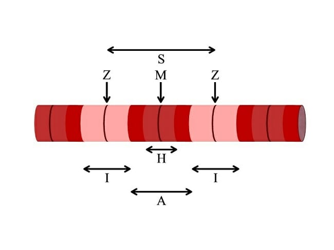
Identify the structures labeled a b and c in the diagram of a sarcomere above. A b and c above. Label the thick and thin filaments in figs. Sign up to access the rest of the document.
In the diagram what is the basic functional unit of a myofibril. Identify the chloroplast structures labeled a b and c. 3 pts this is the end of the preview.
In the diagram below draw three vertical lines showing the locations within a sarcomere of the cross sections indicated by figures a b and c. Many scouting web questions are common. Play games take quizzes print and more with easy notecards.
A actin filament. A sarcomere is the functional unit of striated muscle. 60 the following is a list of the events that occur during a muscle contraction.
Sarcomeres are able to initiate large sweeping movement by contracting in unison. Solved the processes labelled 1 and 2 in the diagram above represent bio 30 high school level science 4 years ago salax0 bioman 5 replies 3130 views. Which of the labeled structures on the diagram holds muscles with similar functions together allows free.
Identify the structures labeled a b and c in the diagram of a sarcomere above. A b b c c f d both b and c e all of these choices are correct. Identify the structures labeled a b c and d 3 pts each a.
In the diagram one of the components of a myofibril is the structure labeled. E multi unit smooth muscle. This means it is the most basic unit that makes up our skeletal muscle.
A b and c above. Herein lies the sarcomeres main purpose. A g b h c b d j e none of these choices are correct.
B and c in the diagram of a sarcomere above. The light dependent reactions occur in the thylakoids. This flashcard is meant to be used for studying quizzing and learning new information.
Skeletal muscle is the muscle type that initiates all of our voluntary movement. Identify the labelled structures a and b in the diagram. In the diagram below draw three vertical lines showing the locations within a sarcomere of the cross sections indicated by figures a b and c.
Label the thick and thin filaments in figs. B myosin filament. A k b i c l d m e a 56.
Label each of the lines. Section 82 end of chapter questions study guide by fmoran29 includes 5 questions covering vocabulary terms and more. The diagrams in model 3 are cross sections of a sarcomere that show the filaments at various locations within a sarcomere.
C is ovarian tissue. Study multi choice chapter 10. In which structures do the light dependent reactions occur.
 Solved 4 Identify The Structures Labeled A And B On The
 Molecular Structure Of The Sarcomeric Z Disk Two Types Of Titin
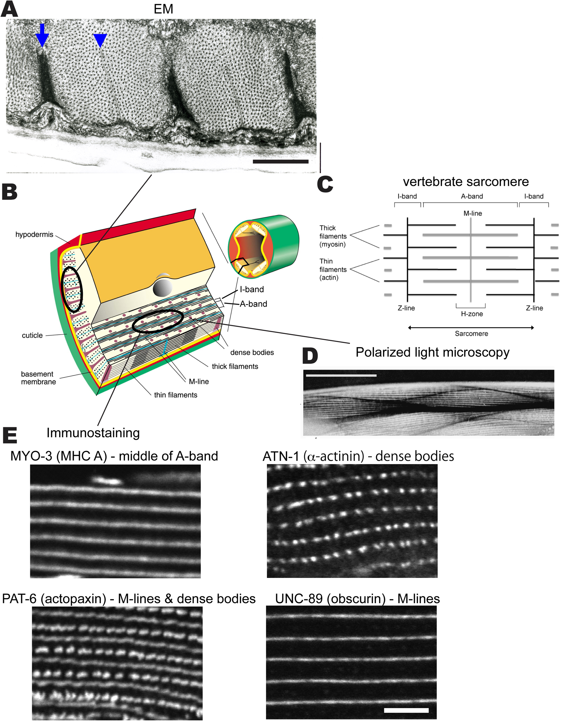 Development Structure And Maintenance Of C Elegans Body Wall Muscle
 Sarcomere An Overview Sciencedirect Topics
 Structure And Function Of Skeletal Muscle
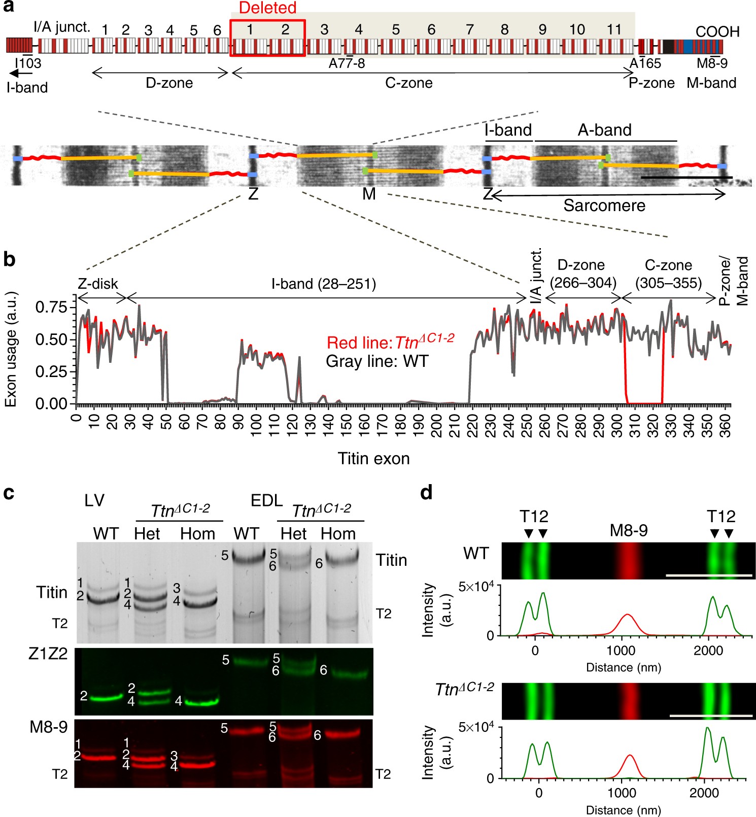 The Giant Protein Titin Regulates The Length Of The Striated Muscle
 Multi Choice Chapter 10 Muscle Tissue Flashcards Easy Notecards
Solved Question 4 Shown Below Are 2 B Lymphocytes I Iden
 A
A
 Associate Degree Nursing Physiology Review
 Muscle Celebrate Cytochemistry Gwen V Childs Ph D
 Ordering Of Myosin Ii Filaments Driven By Mechanical Forces
 10 2 Skeletal Muscle Anatomy Physiology
 Muscle Tissue Junqueira S Basic Histology Text And Atlas 15e
 Sarcomere Wikipedia
 Skeletal Muscle Physiology
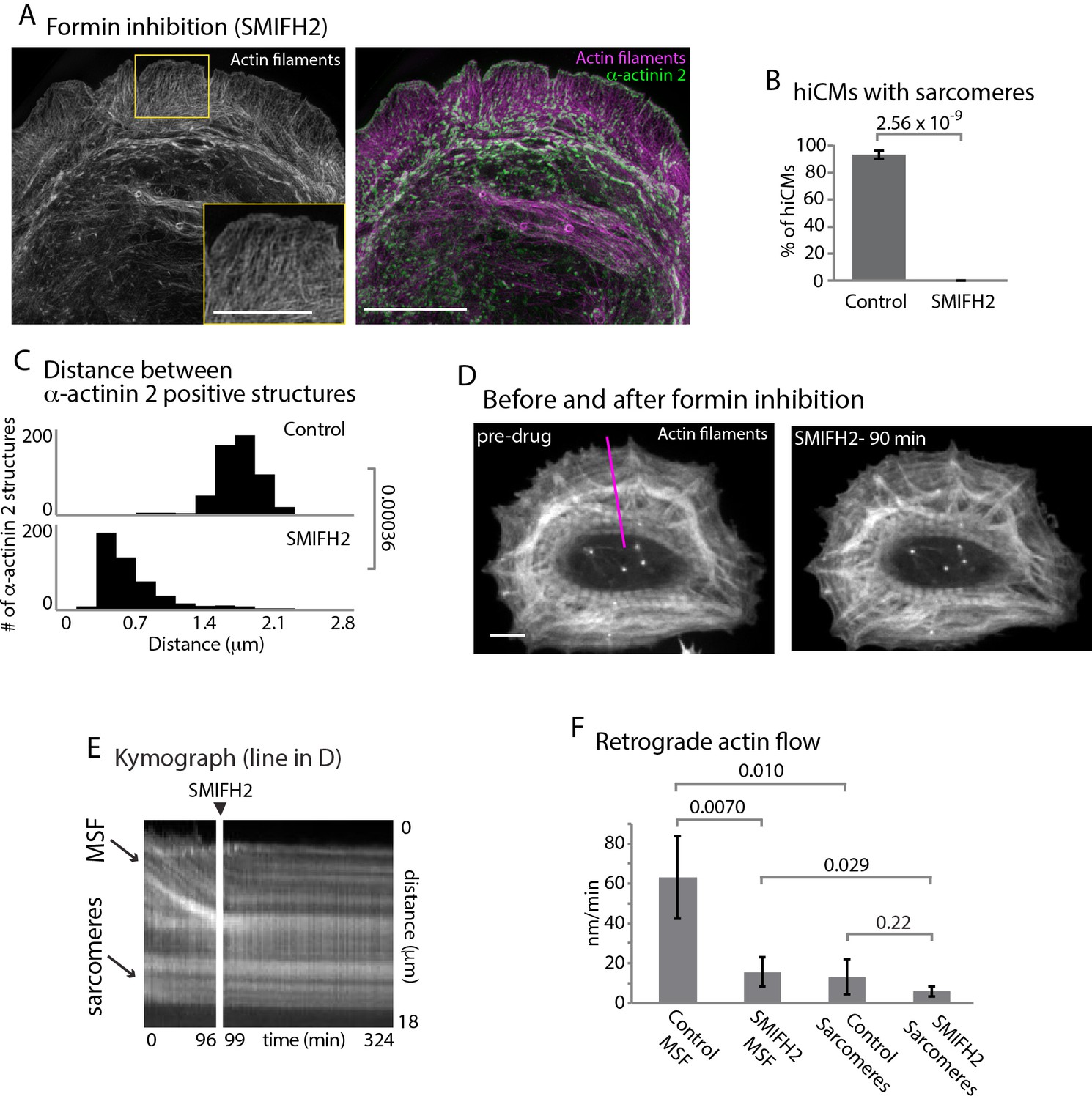 Muscle Specific Stress Fibers Give Rise To Sarcomeres In
 11 2 Muscles And Movement Bioninja
 2 The Sarcomere Length Tension Relation
 Sarcomere Definition Structure Function And Quiz Biology


0 Response to "Identify The Structures Labeled A B And C In The Diagram Of A Sarcomere Above"
Post a Comment