In The Diagram Where Is The Osteon
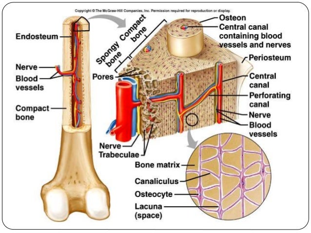
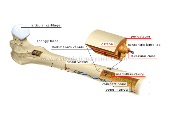
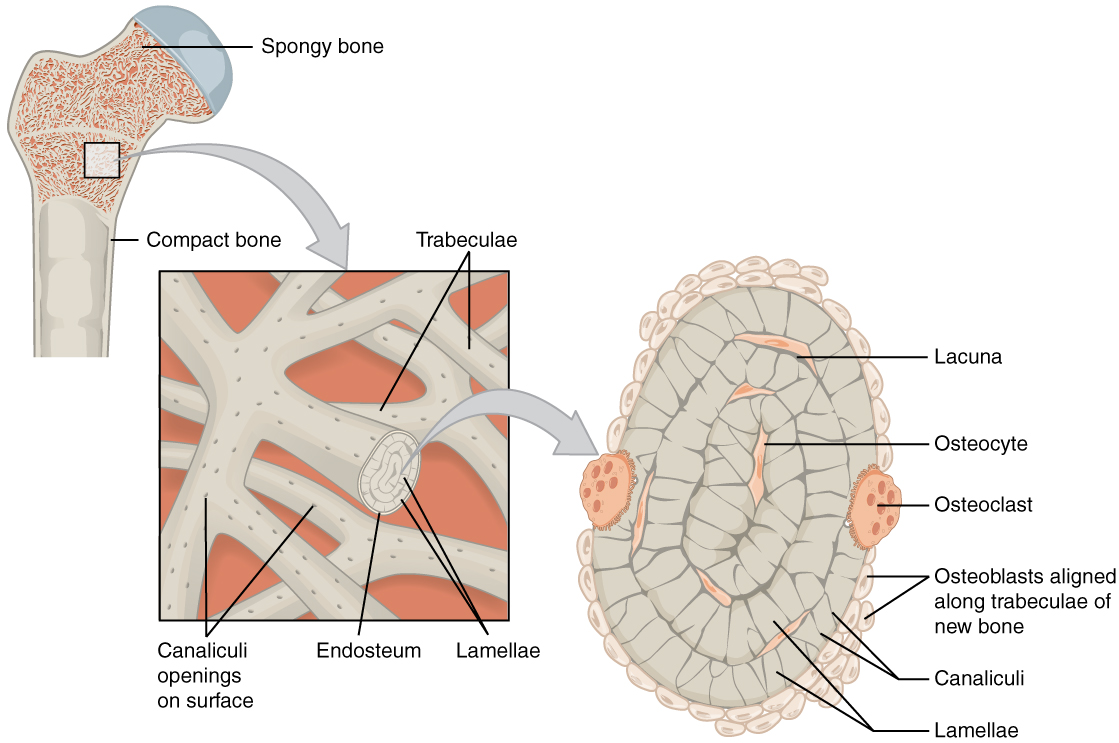
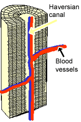
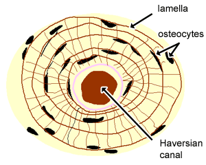
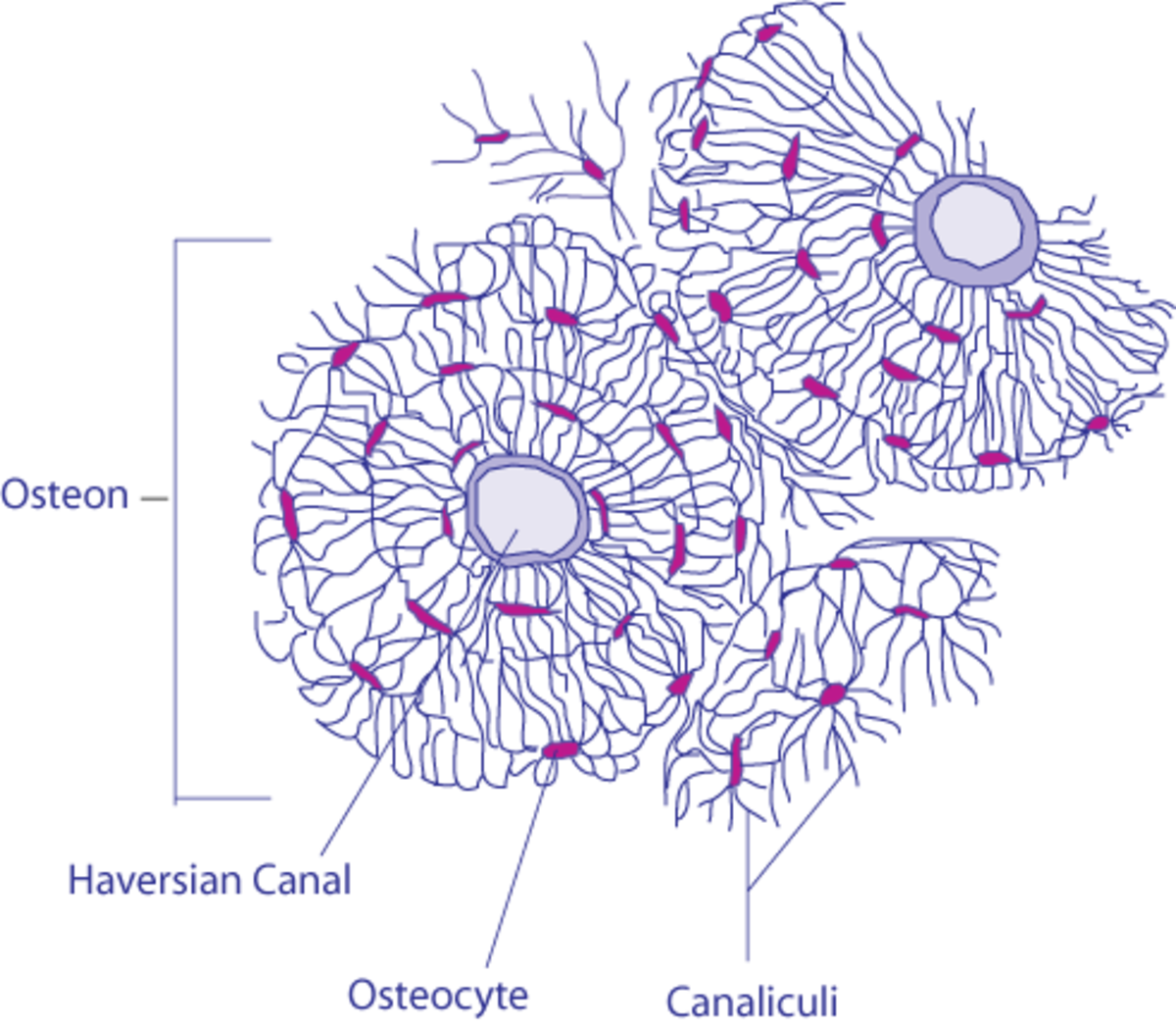
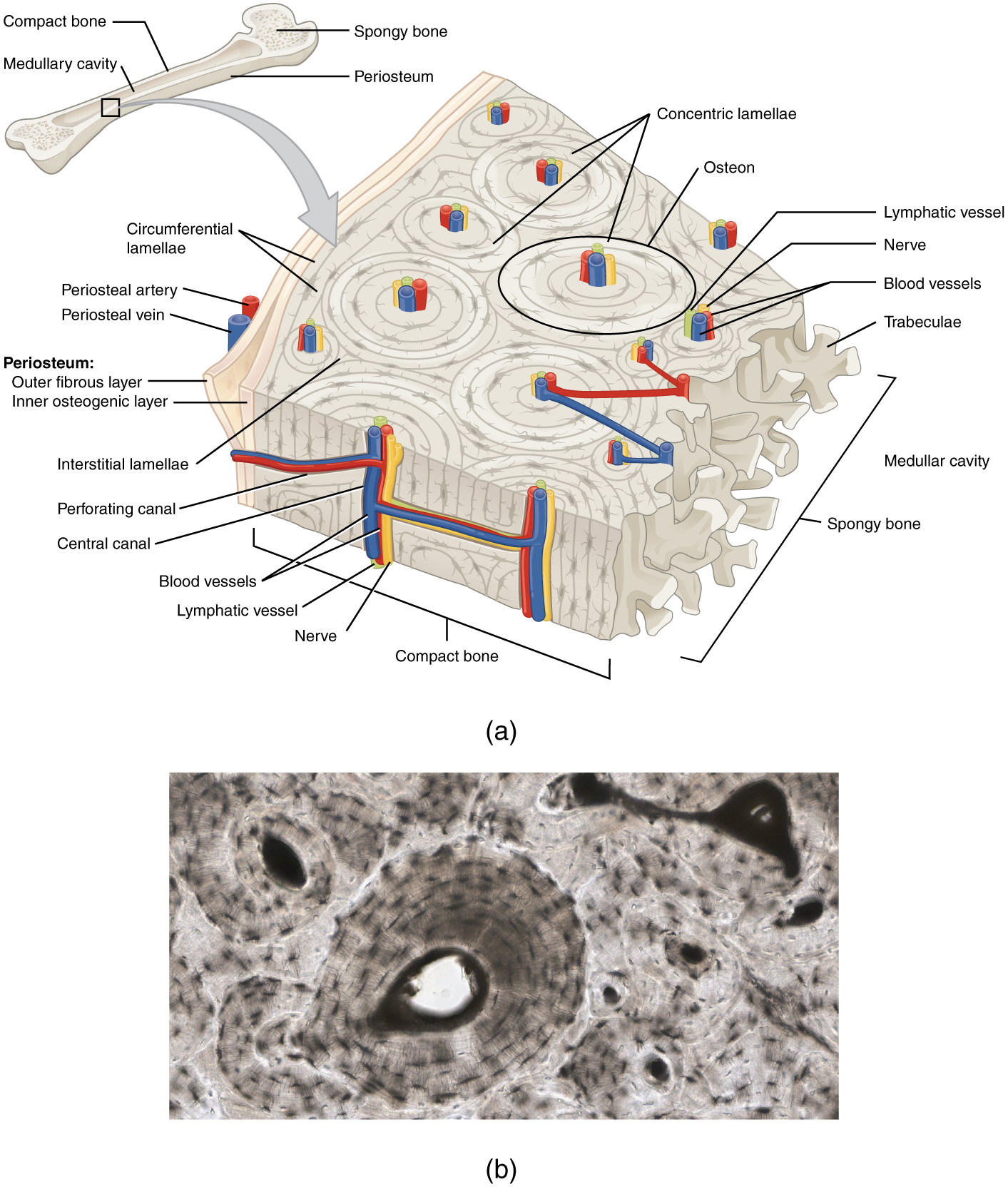
633 compare the structural and functional differences between compact and spongy bone tissue. Each osteon consists of concentric layers or lamellae of compact bone tissue that surround a central canal the haversian canal.
Start studying osteon and other label stuff.

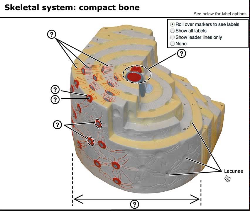
In the diagram where is the osteon. The osteons are closely packed with osteocytes lined up in concentric rings. Osteons roughly cylindrical structures that are typically several millimeters long and around 02mm in diameter are present in many of the bones of most mammals birds reptiles and amphibians. Easy learning objective 1.
Compactness of the bone. The space between the osteons the glue that holds the fibers together. Bony lamella that encircles the outer or inner surface of a bone.
41 in the diagram where is the osteon. Functional unit of the skeletal system. Were the marrow is located basic unit of structure in compact bone comprised of lamellae central canal and osteocytes.
The boundary of an osteon is the cement line. Log in sign up. Osteon and other label stuff.
Log in sign up. Learn vocabulary terms and more with flashcards games and other study tools. Structure and function of the haversian system explained with diagrams.
This imparts a hard and dense texture to the compact bones. Each haversian canal is surrounded by varying number 5 20 of concentrically arranged lamellae of bone matrix. The terms haversian system or osteon refer to the basic cylindrical shaped structural unit of a compact bone which in turn forms a substantial part of the structure of the long bones of the human body.
In the diagram where is the osteon. The haversian canal contains the bones blood supplies. The osteon or haversian system is the fundamental functional unit of much compact bone.
63 histology of bone tissue. 63 describe and compare the properties of compact and spongy bone tissue. A thin layer membrane scale or platelike tissue or part especially in bone tissue.
Layers on bone tissue found in compact bone lamellae osteocyte central canal osteon part of the a bone cell forms when an osteoblast becomes embedded in the.

 Development Structure And Organization Of Bone
 Osteon Of Human Bone Youtube
 This Photo Shows A Model Of An Osteon It Points Out The Blood
Differentiating Fragmented Human And Nonhuman Long Bone Using Osteon
 6 3 Bone Structure Anatomy And Physiology
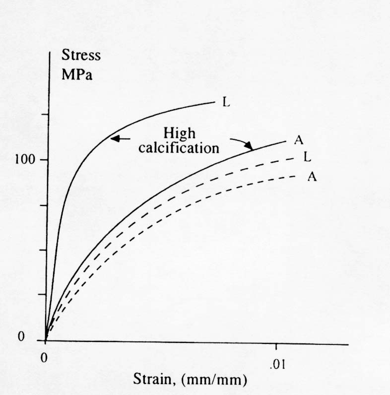
Frontiers In Bioscience 17 1551 1581 January 1 2012 1551 The
![]() Osteon Wikipedia
Osteon Wikipedia
 Bone Structure Anatomy And Physiology I
Vascular Spaces In Compact Bone
 The Histology Guide Cartilage Bone Ossification
 The Histology Guide Cartilage Bone Ossification
 Structure And Function Of The Haversian System Explained With Diagrams
 Copyright C John Wiley Sons Inc All Rights Reserved Chapters 6
 Structure Of Bones Biology For Majors Ii
 Osteoblasts Osteoclasts Calcium And Bone Remodeling Owlcation
 Haversian Canal An Overview Sciencedirect Topics
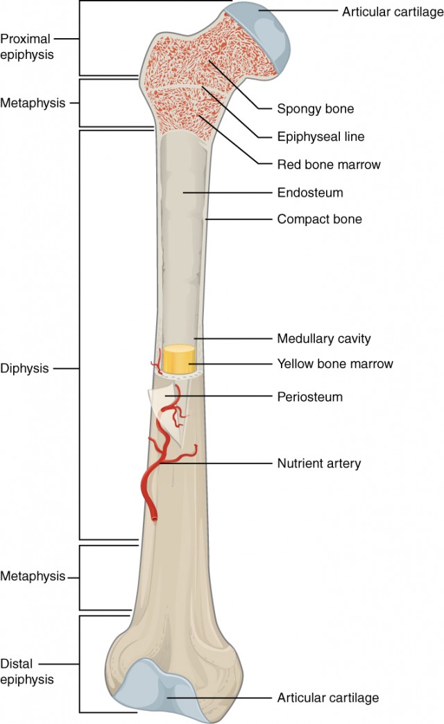
 In This Diagram Of A Long Bone Which Type Of Bone Marking Is

The Skeletal System Ck 12 Foundation
 Lamellae Diagram Wiring Diagram
 6 3 Bone Structure Anatomy And Physiology
0 Response to "In The Diagram Where Is The Osteon"
Post a Comment