Drag The Labels Onto The Diagram To Identify The Parts Of A Knee Jerk Reflex
Muscle reflexes click on the link or the image below for an interactive concept map activity then answer the questions to the right. Label the components of a knee jerk reflex part a drag the labels onto the diagram to identify the parts of a knee jerk reflex.
 The Peripheral Nervous System And Reflex Activity
Although the central pathway involve only spinal cord segment control.

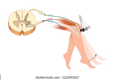
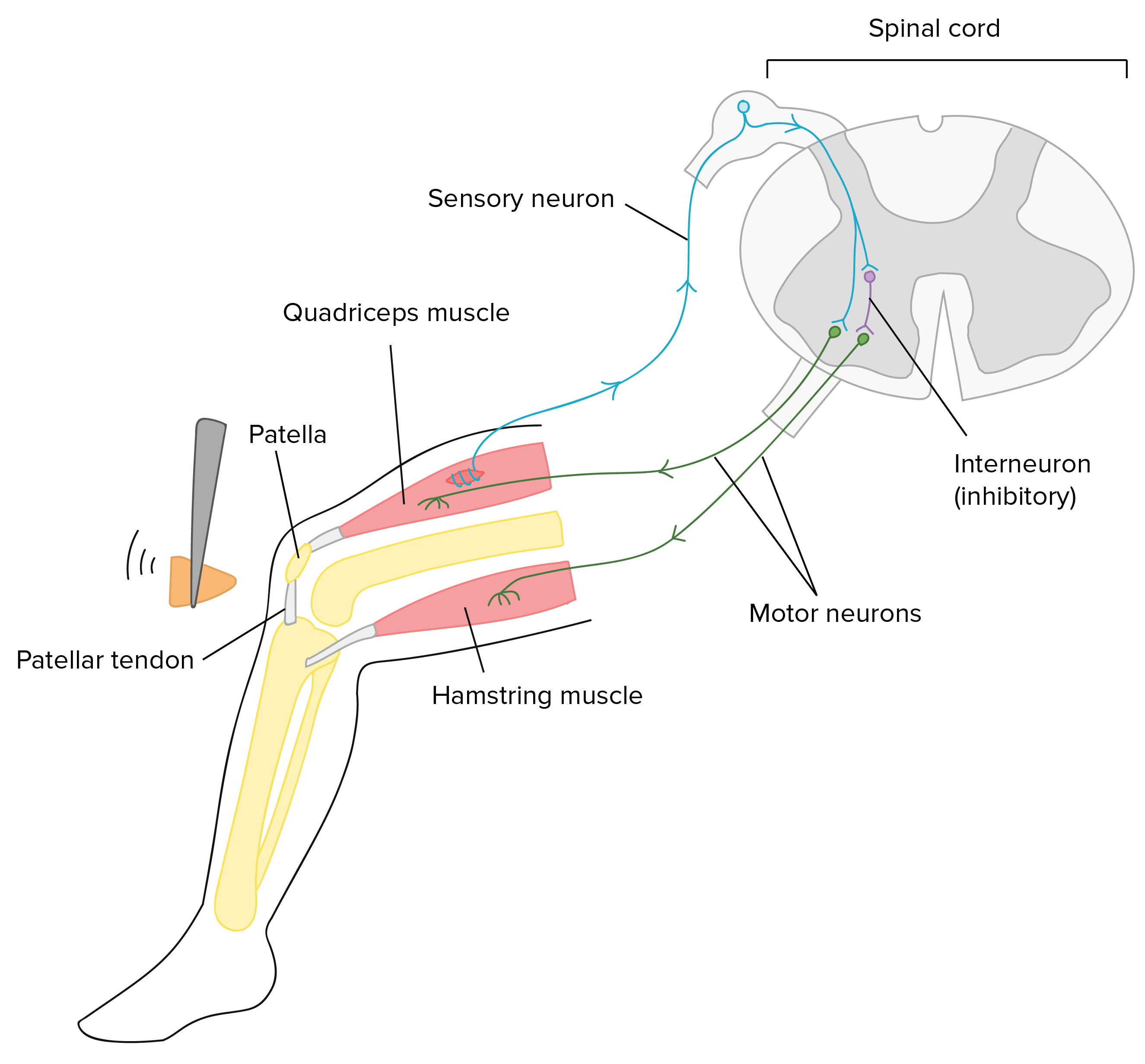
Drag the labels onto the diagram to identify the parts of a knee jerk reflex. Spinal reflex the inborn reflexes mediated by control centers in the spinal cord. Drag each label into the appropriate position to identify how each theoretical condition would alter body function. Label the parts of a monosynaptic reflex arc.
The frontal section is on the right. Part a drag the labels onto the diagram to identify the parts of a kneejerk reflex. The patellar tendon k monosynaptic activation of somatic motor neuron sensory neuron an action potential muscle spindle stretched and effector contracts stretch sensitive neurons are activated inhibited motor neuron does not activate antagonistic muscle response extension of the leg inhibitory interneuron.
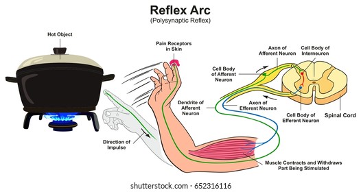
For example when one foot steps on a nail the crossed extensor reflex shifts the bodys weight onto the other foot protecting and withdrawing the foot on the nail. The pathway usually involve cranial and cervical spinal nerves. Drag the labels onto the diagram to identify the parts of a knee jerk reflex.
Reflexes or reflex actions are involuntary almost instantaneous movements in response to a specific stimulus. Learn vocabulary terms and more with flashcards games and other study tools. Cranial reflexes the inborn reflexes by control centers in the brain.
Part a drag the labels onto the diagram to identify the parts of a monosynaptic reflex arc. The more complete diagram of body cavities is provided at the bottom as a reminder of the larger relationships. Epimysium covers whole muscle allows muscles to contract with respect to each other without friction between them perimysium covers the fascicles endomysium covers each muscle fiber tendons formed by the fusion of these 3 connective tissues.
To answer the question you need only name the lungs diaphragm and intercostal muscles parietal and visceral pleural membranes and the pleural cavities. Label the components of a knee jerk reflex part a drag the labels onto the diagram to identify th. Part a drag the labels onto the diagram to identify the parts of a knee jerk reflex.
 Human Biology Ap Biology Science Khan Academy
 Nervous System Anatomy
 Reflex Images Stock Photos Vectors Shutterstock
 14 5 Sensory And Motor Pathways Anatomy Physiology
 Human Physiology Print Version Wikibooks Open Books For An Open World
 Chapter 13 Flashcards Easy Notecards
 Nervous System Anatomy
 Lab Final Systems Physiological Science Bi315 With Widamier At
 Section I Mobility And Musculoskeletal System Hazzard S Geriatric
 A P Chapter 11 Nervous System 2 Homework Example Graduateway
 Human Physiology Print Version Wikibooks Open Books For An Open World
 Reflexes Images Stock Photos Vectors Shutterstock
 Motor Disorders Clinical Neurology 9e Accessmedicine Mcgraw
Sense Organs Handout
 Human Biology Ap Biology Science Khan Academy
 Patella Images Stock Photos Vectors Shutterstock
 Nervous System Anatomy
 1 4 The Somatic Nervous System Neuroscience Canadian 1st Edition
 14 3 The Brain And Spinal Cord Anatomy Physiology
The Peripheral Nervous System And Reflex Activity
 Reflex Images Stock Photos Vectors Shutterstock
 Human Biology Ap Biology Science Khan Academy
 1 4 The Somatic Nervous System Neuroscience Canadian 1st Edition
Antphy 211 Study Guide 2013 14 Whitehead Instructor Whitehead At

0 Response to "Drag The Labels Onto The Diagram To Identify The Parts Of A Knee Jerk Reflex"
Post a Comment