In The Diagram Of Skin Shown Below Which Labeled Structure Generates Fingerprints
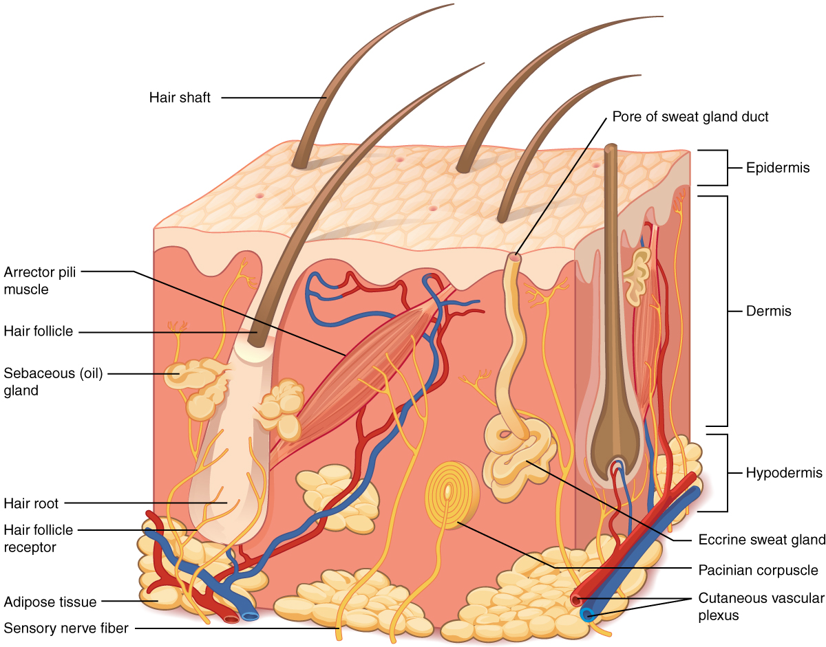
Collagen fibers become more organized fibroblasts. Oil and sweat secretions leave the marks of these ridges on smooth surfaces thus leaving a fingerprint.
Osa Optical Fingerprint Recognition Based On Local Minutiae
These ridges have a unique shape that can be used to identify people.
In the diagram of skin shown below which labeled structure generates fingerprints. Asked sep 20 2015 in anatomy physiology by rubyor. A a b b c d d e e f. The ducts of sweat glands open on the tops of the epidermal ridges as sweat pores the sweat and ridges form fingerprints or footprints on touching a smooth object.
The layer of skin that forms thefingerprint is the epidermis. 51 describe the composition of the epidermis and dermis. Describe the structure and function of the different types of exocrine glands found in the skin.
Answered sep 20 2015 by. Fingerprints are produced by the epidermis. A sequence of amino acids linked by peptide bonds.
In the diagram of skin shown below which labeled structure in the diagram of skin shown below which labeled structure generates fingerprints pictures in the diagram of skin shown below which labeled structure download figure open in new tab. In the diagram of skin shown below which labeled structure generates fingerprints. 28 in the diagram of skin shown below which labeled structure generates fingerprints.
Describe how fingerprints are formed and what they are used for. Medium learning objective 1. In the photomicrograph of a portion of thick skin shown below which layer is the stratum basale.
The fingerprint is an impression or mark on the fingertip thatuniquely identifies an individual. So 511 describe the layers of the epidermis and the cells that compose them. So 51 describe the composition of the epidermis and dermis.
Contains information necessary for the protein to fold into its higher order structures. A a b b c g d h e none of these answer choices are correct. The carbonyl oxygen is positioned on one side of the c n bonds and the amino hydrogen is on the opposite side.
Medium study objective 1. In the diagram of skin shown below where is the reticular region of the dermis. The primary structure of a protein consists of.
They are caused by the friction ridges on the outermost layer of the skin. In the diagram of skin shown below which labeled structure generates fingerprints. Does not contain hair follicles.
Contains more sweat glands than thin skin. Found in the palms soles of the feet and fingertips.
 Clinical Exome Sequencing Reveals Locus Heterogeneity And Phenotypic
Proteomic Analysis Of Altered Protein Expression In Skeletal Muscle
The Effects Of Plant Flavonoids On Mammalian Cells Implications For
 Intelligent Fingerprint Quality Analysis Using Online Sequential
 50 Years Of Biometric Research Accomplishments Challenges And
 Molecular Evidence Of Burkholderia Pseudomallei Genotypes Based On
 Raman Spectroscopy With A 1064 Nm Wavelength Laser As A Potential
 Ep2532353a1 Herbal Therapy For The Treatment Of Asthma Google
Global Transcriptome Analysis And Enhancer Landscape Of Human
 Transcriptional Fingerprints Of Antigen Presenting Cell Subsets In
Osa Dual Beam Manually Actuated Distortion Corrected Imaging Dmdi
 Ep2253719a2 Diagnosing And Monitoring Transplant Rejection Based
 Cs379c 2018 Class Discussion Notes
 Question Type Multiple Choice 25 A Raised Scar That Extends Into
Mas And Its Related G Protein Coupled Receptors Mrgprs
 What Is A Dna Fingerprint Facts Yourgenome Org
 The Chemical Fingerprint Of Hair Melanosomes By Infrared Nano
A Novel Dnmt3b Splice Variant Expressed In Tumor And Pluripotent
 5 1 Layers Of The Skin Anatomy And Physiology
 12 2 Visualizing And Characterizing Dna Biology Libretexts
 Ijms December 2018 Browse Articles
When Whorls Collide The Development Of Hair Patterns In Frizzled 6
0 Response to "In The Diagram Of Skin Shown Below Which Labeled Structure Generates Fingerprints"
Post a Comment