Human Cell Diagram To Label
The outer covering of the cell is known as cell membrane. This happens because the cells take the normal cell cycle through copying and separation of chromosomes but do not go the whole way and divide.
 Blank Human Cell Diagram To Label Diagram
Blank animal cell diagram worksheet.

Human cell diagram to label. Use the word bank below to identify the parts of the human cell. Other sets by this creator. Label 5 parts of the human cell.
The first is a colored and labeled cell diagram. They form by fusion of cells with one nucleus. Name three of the images in the following diagram.
Human body label the part of the human body. The third and fourth diagrams are animal cell diagram worksheets. Human cell diagram school science projects science lessons life science science week printable crafts free printable printables preschool science.
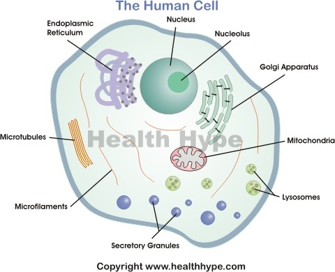
The cell membrane is the outer coating of the cell and contains the cytoplasm. Quiz yourself by filling in the blanks. The lipid molecules on the outer and inner part lipid bilayer allow it to selectively transport substances in and out of the cell.
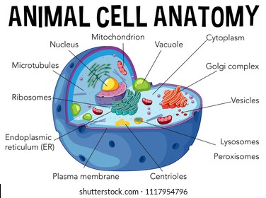
Can you name the different parts of an animal cell. Test your knowledge on this science quiz to see how you do and compare your score to others. The next is a black and white version of the first.
The endoplasmic reticulum er is a membranous structure that contains a network of tubules and vesicles. Log in sign up. Human cell diagram parts pictures structure and functions cell membrane.
The cell is the basic unit of the human body. Cells group together to make skin bones and blood. Cytoplasm is the name given to the filling fluid of the cell which is also known as.
Anatomy of human cell diagrams. Anatomy of a human cell learn with flashcards games and more for free. Human cell properties diagram parts pictures structure cell membrane.
Skeletal muscles such as your biceps have very long cells with many nuclei. Label 5 of parts on the following diagram. Animal cell anatomy label the animal cell diagram using the glossary of animal cell terms.
Inside the cell nucleus is dna which identifies the color of hair eyes and skin. Some cells in adult heart muscle have two nuclei. Human skeleton label the major bones in this human skeleton printout.
There are over one billion cells in each human body. Log in sign up. Endoplasmic reticulum or er is made of.
Anatomy final exam 12. The arm in english label the parts of the arm and hand in english. Labeled animal cell diagram.
It is a double layered membrane composed of proteins and lipids. The golgi apparatus is a stacked collection of flat vesicles.
 Human Cells To Label Worksheet By Jklawrence Teaching Resources Tes
 6th Malkowski L Science Ela Vocabulary The Cell
 Human Cell Diagram Parts Pictures Structure And Functions
 Human Cell Coloring Page Plant Cell Coloring Pages Human Page With
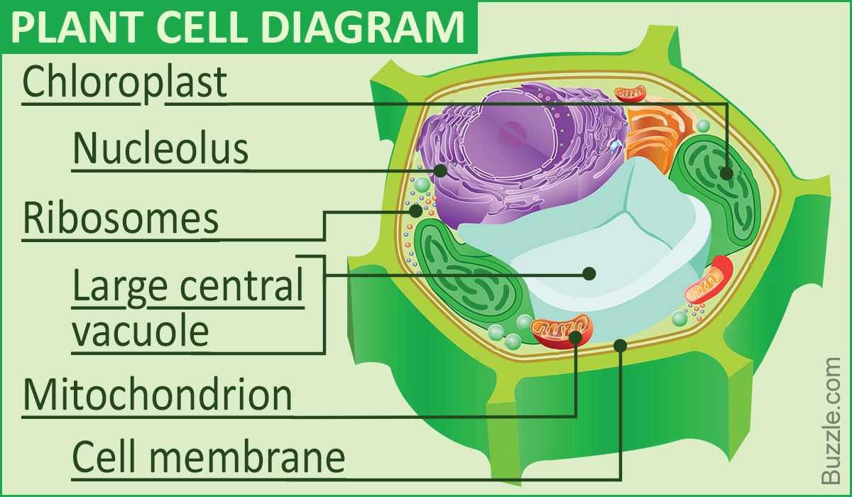
 Plant Diagram To Label Wiring Diagram Database
 Overview Of Neuron Structure And Function Article Khan Academy
 Animal Cell Diagram With Labels Www Topsimages Com
 Printable Cell Structure Diagram Wiring Diagram
 Label Animal Cell Worksheet The Best Worksheets Image Collection
 Diagram Of Brain Tips For Using Worksheets Human Cell To Label
 Human Cell Diagram To Label Animal Cell Biology Pictures Animal
 Real Cell Diagram 15 16 Stromoeko De
 Chromatin Capture Links The Metabolic Enzyme Ahcy To Stem Cell
 A Animal Cell Diagram 1 Wiring Diagram Source
 Human Physiology Cell Structure And Function
Human Anatomy
 Leaf Cell Diagram Label Wiring Schematic Diagram
 Skin Cell Diagram Labeled Michaelhannan Co
 Basic Plant Cell Diagram With Labels New Human Cell Diagram Parts
Label The Human Cell By Caroline Mckeever Teaching Resources Tes
 Diagram Of The Heart Labeled Label Muscles Worksheet Body Worksheets
 Label Plant Cell Worksheet The Best Worksheets Image Collection

0 Response to "Human Cell Diagram To Label"
Post a Comment