In The Diagram Where Is The Mastoid Process
It is also filled with sinuses or mastoid cells. The air cells are lined by a thin mucous membrane.
 Mastoid Process Anatomy Pictures And Information
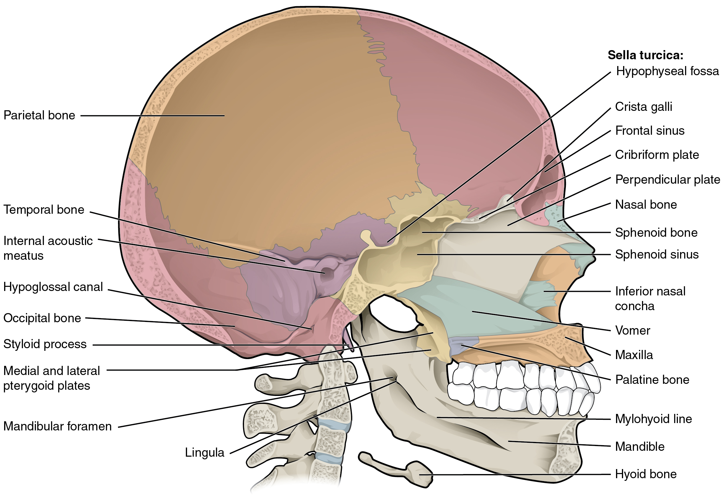
Parts of the frontal and sphenoid bones comprise the roof of the orbit.
In the diagram where is the mastoid process. Ear infections linked to mastoid cells is typically treated with antibiotics. The mastoid is connected to the part of the ear where the hearing and balance mechanisms are located. Small fragile bone making up part of the front inner walls of each eye socket and providing room for the passage of the lacrimal ducts.
The mastoid is a honeycomb of air cells located behind the ear. Parts of the maxilla zygomatic and palatine bones make up the floor of the orbit. The mastoid process is a point of attachment for the sternocleidomastoid muscles of the neck.
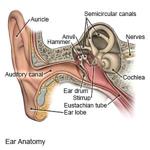
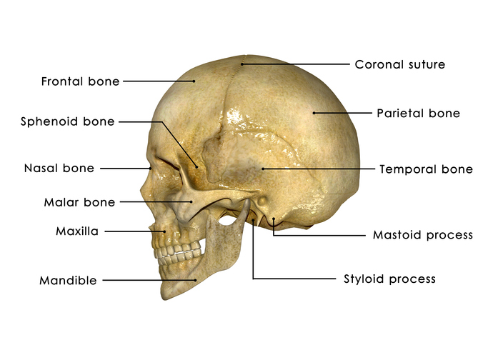
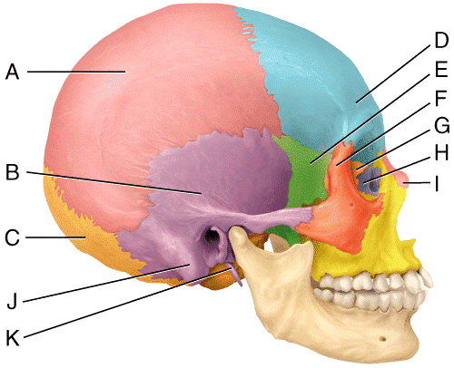
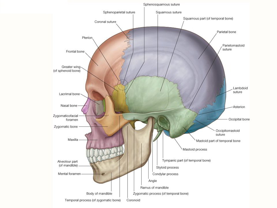
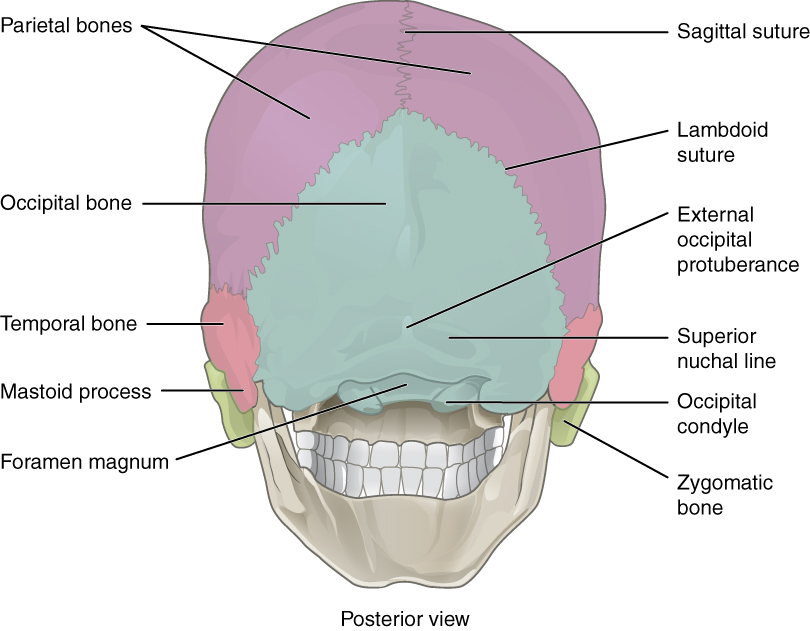
The mastoid process is part of the temporal bone the large bone that runs along the middle bottom of the skull. The mastoid notch is located medial to the mastoid process. You can locate your mastoid if you place your fingers behind your earlobe.
Parts of the zygomatic and sphenoid bones form the lateral wall of the orbit. There is a deep groove on the medial side of the process as well as a shallow furrow called occipital groove which houses the occipital artery. The mastoid process serves for the attachment of the sternocleidomastoid the posterior belly of the digastric muscle splenius capitis and longissimus capitis.
The mastoid process is located just behind the ear in humans. The mastoid process is a small triangular shaped bone that protrudes from either side at the base of your skull. This article covers the anatomy function muscle attachments and clinical aspects of the mastoid process.
The mastoid can be affected by diseases such as infection and cholesteatoma. Click now to learn more at kenhub. Similarly a curved groove known as sigmoid sulcus is present on the inner surface of the bony structure to lodge a part of the transverse sinus.
This article covers the anatomy of the temporal bone its parts connecting sutures and foramina. The main function of the mastoid process is to connect your neck muscles to your skull and help regulate pressure in your ear. The mastoid process lying in the mastoid part of the temporal bone in the human skull is a conicalpyramidal projection present each side of the head at the base of the skull.
Mastoid notch serves as the site of muscle attachment for the anterior and posterior bellies of the digastrics whose function is to open the mouth 2. It is pierced by stylomastoid foramen in front and mastoid foramen is found on its back. The mastoid process is located just behind the ear canal and lateral to the styloid process.
Structure and landmarks of the temporal bone. It is the prominent bony protrusion easily seen behind the earlobes. The bone which forms part of the hard palate of the mouth part of the nasal cavity and part of the orbital cavities.
Parts of the maxilla lacrimal ethmoid and sphenoid bones form the medial wall of the orbit.
 The Skull Anatomy And Physiology I
 Mastoid Process Stock Photos Mastoid Process Stock Images Alamy
 Mastoid Foramen Wikipedia
 Treatment Common Causes Of Jaw Pain Lock Jaw Cpr
 Skull Fractures Parents Accused
 Mastoid Emissary Vein Operative Neurosurgery
Mastoiditis What You Need To Know
 Sg Face Deep
 Mastoid Bone Picture Human Anatomy Study Pinterest Anatomy
 Head Bone Diagram Wiring Schematic Diagram
 The Skull Anatomy And Physiology I
 Print Multi Choice The Skeletal System The Axial Skeleton
 Mastoid Part Of The Temporal Bone Howling Pixel
 Print Multi Choice The Skeletal System The Axial Skeleton
 Mastoiditis What Is It Symptoms Causes Prevention And Treatment
 7 2 The Skull Anatomy And Physiology
 Prominent Ear Correction Otoplasty The Procedure And Its Results
 Sugar Skull Planning
 Easy Notes On Norma Lateralis Learn In Just 4 Minutes Earth S Lab
 7 2 The Skull Anatomy And Physiology
 Diffrentiate Male Skull From Female Skull
 Erector Spinae Muscle Consists Of 3 Muscles Iliocostalis
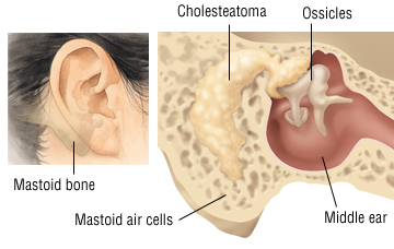 Chronic Otitis Media Cholesteatoma And Mastoiditis Harvard Health
 This Exhibit Depicts The Anatomy Of The Inferior Skull Including


0 Response to "In The Diagram Where Is The Mastoid Process"
Post a Comment