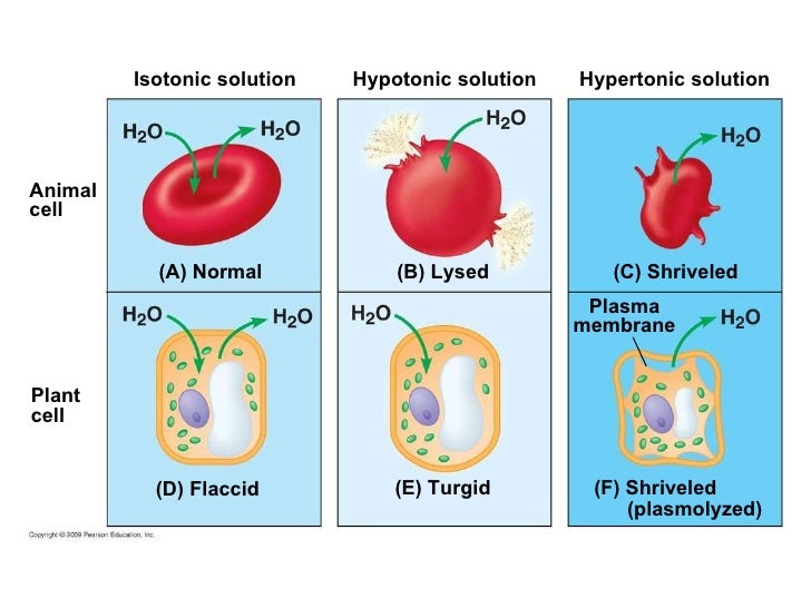
In The Diagram Which One Represents A Hypertonic Solution
Find your answers solutions and more. Step four the amino acid on the trna at p site forms a peptide bond with the amino acid at a site.
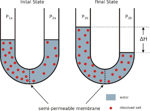
 13 10 Osmosis Why Drinking Salt Water Causes Dehydration
In the diagram shown below which of the indicated structures is composed of basal lamina and.

In the diagram which one represents a hypertonic solution. Step five the trna at the p site leaves the ribosome and the ribosome shifts down by one codon. In the diagram which one represents a hypertonic solution anatomy and physiology which of the light micrographs in the figure below shows a stratified cuboidal epithelium. In this type of transport process a solute eg.
What structural component of the membrane is labeled e in the diagram. Guidelines 1 to 5 indicate the following. In the diagram which one represents a hypertonic solution anatomy and physiology.
We made it much easier for you to find exactly what youre looking for on sciemce. In the diagram this structure directs cellular activities. Anatomy and physiology.
Guidelines 1 to 5 indicate the following. In the diagram which one represents a hypertonic solution. Where on the diagram is the femoral area.
A a b b c c d both b and c e. Easy study objective 1. Enjoy our search engine clutch in the diagram which one represents a hypertonic solution.
Asked sep 19 2015 in anatomy physiology by wayuvan. The trna previously at the a site is now at the p site. Help and review science courses.
In the diagram which one represents carrier mediated facilitated diffusion. The below diagram represents a plant cell after being placed in a strong sugar solution. In the biological fields this generally refers to a solution that has less solute and more water than another solution.
Lecture quiz practice 1. 52 in the diagram which panel shows the kinetochore of the centromeres aligning along the center of the mitotic spindle of the cell. Which of the following represents a receptor a a b b.
This is an organelle that modifies and stores proteins produced elsewhere. Definition example diagram. Anatomy and physiology.
Glucose binds to a specific carrier protein on one side of the membrane. So 37 understand the events and processes involved in cell division. A hypotonic solution is any solution that has a lower osmotic pressure than another solution.
One type of human cell spermatozoa is capable of movement because it has this type of microtubule. This binding induces a conformational change in the carrier protein that results in the solute moving down its concentration gradient to the other side of the membrane. Anatomy and physiology.
These are small flattened curved membranous sacs with bulging edges. Enjoy our search engine clutch. This organelle contains ribosomes which synthesis proteins.
 3 1 The Cell Membrane Anatomy And Physiology
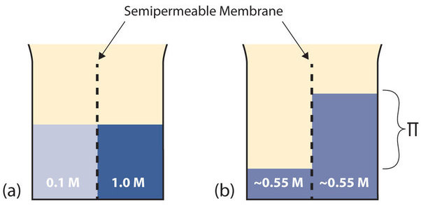
 Osmotic Pressure Wikipedia
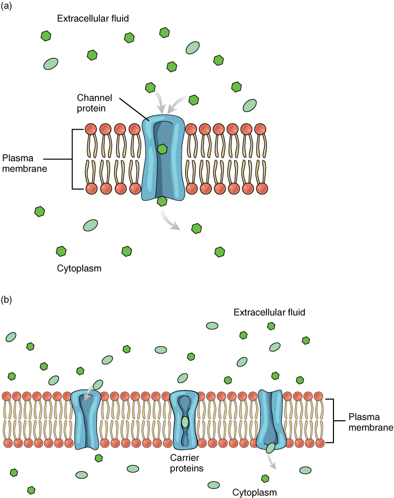
5 2 Passive Transport Texas Gateway

 Solved Select The Diagram That Represents The Shape Of A Red B
 What Happens To A Cell As It Is Placed In A Hypertonic Solution
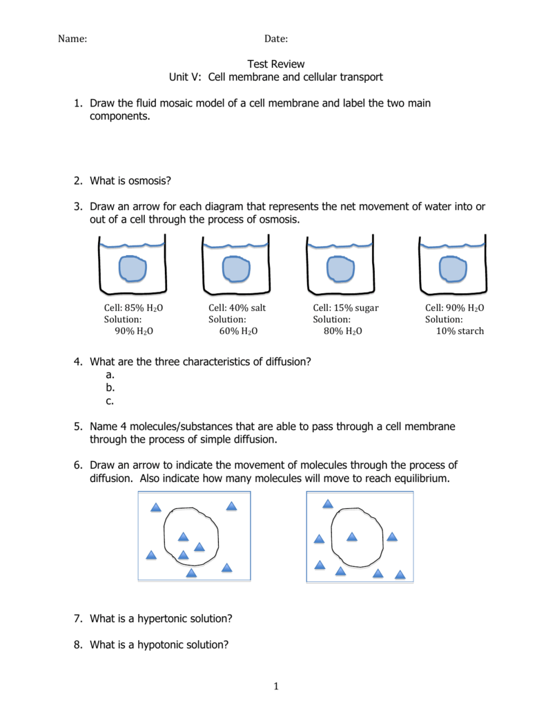 Unit 5 Test Review
Osmotic Behaviour Of Human Mesenchymal Stem Cells Implications For


 Anatomy And Physiology Of Animals The Cell Wikibooks Open Books
 Ca12 E K Mutation Modulates Volume Regulation Of Aqp5 A Volume
 What Would Happen To Your Red Blood Cells If They Were Placed In
Rectification Of The Water Permeability In Cos 7 Cells At 22 10 And 0 C
![]() Hypotonic Solution Induced Ca 2 Transients In Pasmcs Of Normoxic And
Hypotonic Solution Induced Ca 2 Transients In Pasmcs Of Normoxic And
Name Date Period
Hypertonic Solution An Overview Sciencedirect Topics
Lab Exercise 3
 Ch5
Ch5

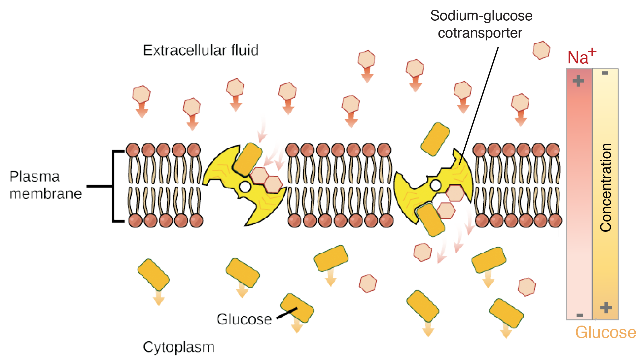 Membranes And Transport Biology Science Khan Academy
0 Response to "In The Diagram Which One Represents A Hypertonic Solution"
Post a Comment