Diagram Of Head And Neck
The neck contains seven of these known as the cervical vertebrae. From supporting the head to containing the spinal cord and nerves as they emerge from the skull this structure does it all.
 Blood Supply Of The Head And Neck Stock Vector Illustration Of
The skeletal section of the head and neck forms the top part of the axial skeleton and is made up of the skull hyoid bone auditory ossicles and cervical spine.

Diagram of head and neck. The spinal column contains about two dozen inter connected oddly shaped bony segments called vertebrae. The human head weighs nearly as much as the average bowling ball at around ten to twelve pounds respectively. Learn vocabulary terms and more with flashcards games and other study tools.
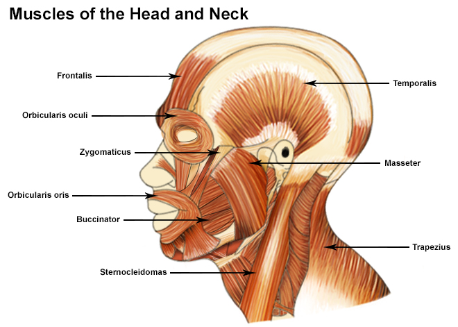
Superficial dissections of the head and neck as seen in the gallery show the many different muscles that are required for movement plus those that control facial expression. Neck anatomy pictures bones muscles nerves. Rotation describes the action of moving the head from side to side lateral motion brings the ear to the shoulder flexion moves the chin to the chest as in looking down.
Rotation lateral flexion flexion and hyperextension. Anatomy and function of the temporal temporalis muscle. Start studying 08 muscles of head and neck.
Origin insertion innervation and functions of the masseter muscle. Learn this topic now at kenhub. The group locations of greatest clinical importance include the following areas namely groin armpit neck under the jaw and chin behind the ears and on the back of the head.
Origin insertion innervation and functions of the lateral pterygoid muscle. The neck is the start of the spinal column and spinal cord. Radiological anatomy of the head and neck on a ct in axial coronal and sagittal sections and on a 3d images.
Anatomy of the head and neck ct scan ct scan of head and neck. The muscle anatomy of the head and neck is a fascinating area with the the neck also containing the 7 vertebrae of the part of the spine called the cervical curve. Major muscles of facial expression and mastication and neck muscles.
Regions of the head and neck this article will discuss the anatomy of the regions of the head and neck and clinical importance. They are the smallest and uppermost vertebrae in the body. Atlas main muscles of the head and neck.
With the exemption of relatively few individual nodes most of the lymph nodes do come together in groups in specific areas of the body. The neck has the ability to support a great deal of weight too. The motion of the muscles of the neck are divided into four categories.
The head is rests on the top part of the vertebral column with the skull joining at c1 the first cervical vertebra known as the atlas.

 Seer Training Muscles Of The Head And Neck
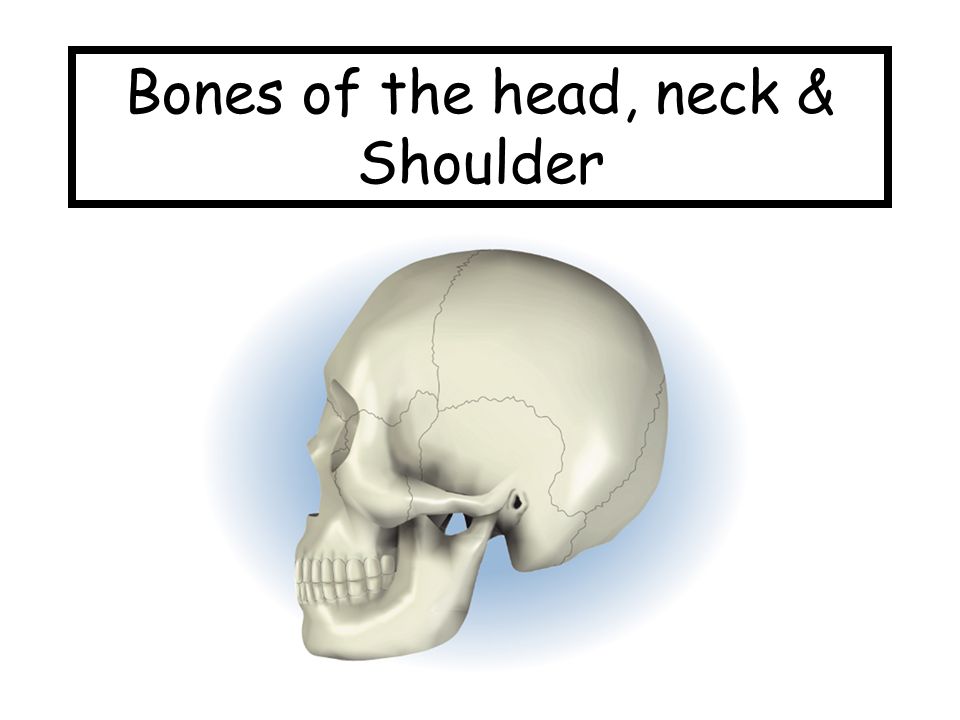
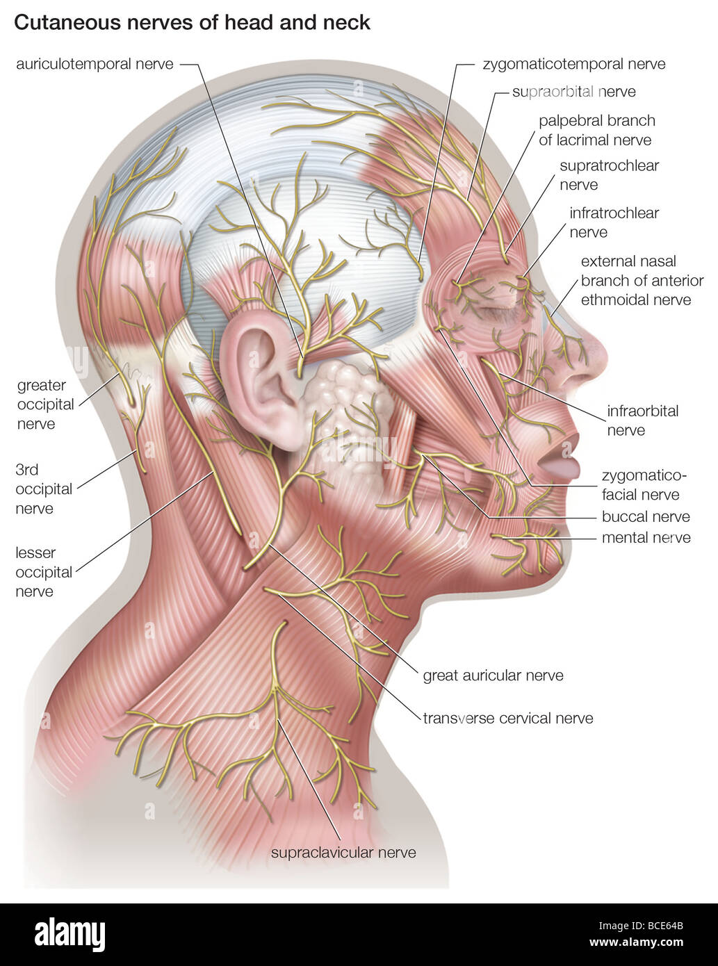 Diagram Of The Cutaneous Nerves Of The Head And Neck Stock Photo
 So Many Muscles That Cause Migraines Arm Neck Shoulders And Back
 Pictures Guide To Cancers Of The Head And Neck
 Head And Neck Cancers Cdc
 Ucsd S Practical Guide To Clinical Medicine
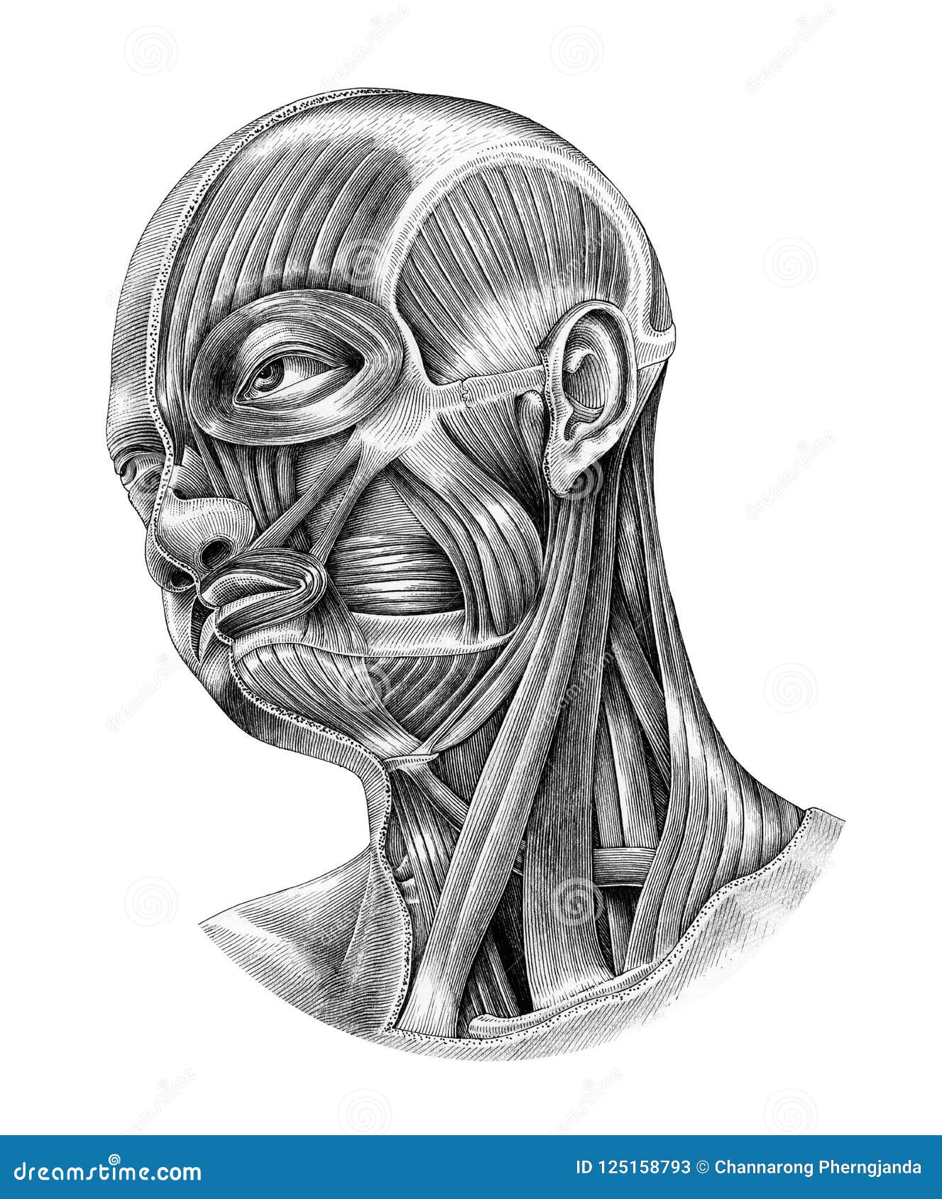
 File Illu Head Neck Muscle Jpg Wikimedia Commons

 Diagram Of Lymph Nodes In Neck Lymphatic System Neck Diagram Lymph

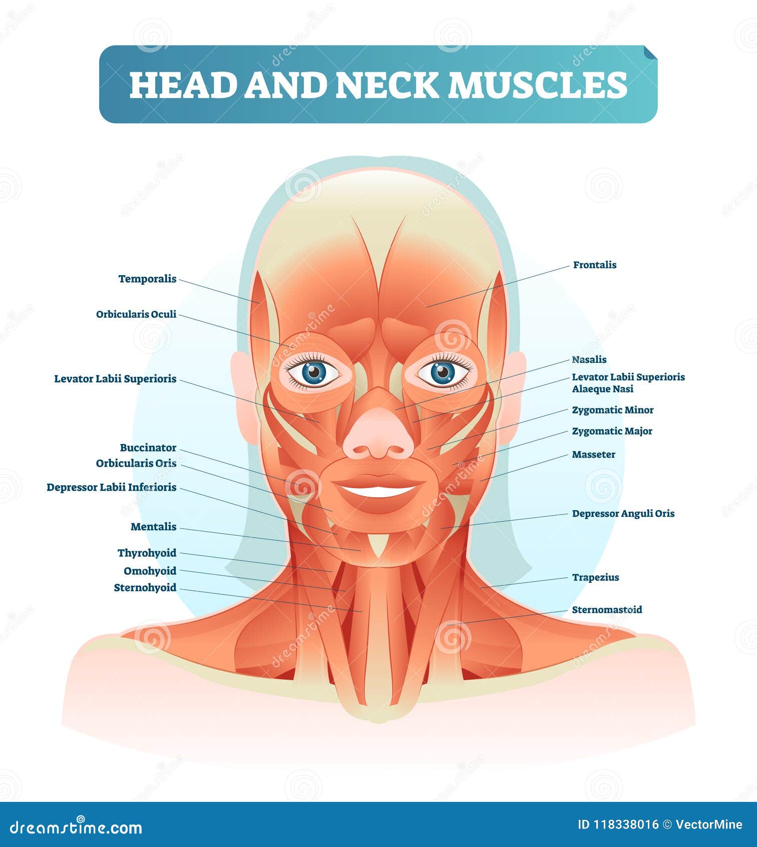 Head And Neck Muscles Labeled Anatomical Diagram Facial Vector
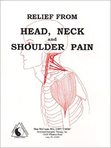 Relief From Head Neck And Shoulder Pain Don Mccann 9780970681119
 Face Wikipedia
 Circulation Head And Neck Arteries Purposegames
Diagram Of Coolant Flow And The Integrated Layers Of Head Neck
 Head Neck Cancers Cancer Council Victoria
 File Head Neck Tracheostomy Jpg Wikimedia Commons
 Human Head And Neck Anatomy Diagram Illustration Vintage Style I
0 Response to "Diagram Of Head And Neck"
Post a Comment